The T Wave on an Ecg Tracing Represents
The T wave on an ECG tracing represents O atrial depolarization. Each of the following changes will result in increased blood.

Pin On All About Nursing Things To Help Me Remember Because My Brain Is So Full That It May Explode
The T wave represents ventricular repolarization.

. The T wave on an ECG tracing representsA. The other main components of an ECG are. The T wave on an ECG tracing represents A atrial depolarization.
BC atrial repolarization happens at the same time as ventricular repolarization during the QRS Complex and ventricular repolarization covers it. What might a longer-than-normal P-R interval indicate. Represents time it takes for signal to travel throughout atria.
Depolarization of the ventricles Incorrect b. What change in polarization is the t waves on an ecg tracing. The T wave on an ECG tracing represents O atrial depolarization.
The P wave the QRS complex and the T wave. The ECG reveals atrial fibrillation with a variable rate between 110 and 130 beatsmin and a 12-lead ECG tracing reveals the same. P wave It represents the depolarization of atria and represents atrial contraction.
However various waveform morphologies may present as an indication of benign or clinically significant injury or insult to the myocardium. Why isnt there a wave on a normal ECG tracing that represents atrial repolarization. The PR interval is usually.
A U wave is sometimes present. What part of the ECG tracing represents the time it takes for the impulse to activate the myocardium to the complete contraction. Understanding the differential diagnosis for T wave discrepancies is crucial to the successful and safe management of various cardiac.
QUESTION 7 The T wave on an ECG tracing represents A atrial repolarization B from BIOLOGY 2458 at University of Texas Arlington. Repolarization of the ventricles begins at the epicardial surface of the ventricles and progresses inwardly through the ventricular walls to the endocardial surface. The T Wave occurs during the last part of the ventricular systole.
Click to see full answer. Asked Dec 2 2021 in Health Professions by Steve. When computing a heart rate from the ECG tracing the nurse counts 15 of the small.
What part of the ECG tracing represents the repolarization of the bundle of His and Purkinje fibers. The normal T-wave. Between the waveforms are the following segments and intervals.
An additional wave the U wave Purkinje repolarization is often visible but not always. EKG Chapter 2 - cardio system. T-Wave is the type of wave that you usually see on the electrocardiogram or ECG.
The T wave of an ECG pattern represents the repolarization of the. What does the T wave of the electrocardiogram ECG represent. Depolarization of the atria c.
The T wave on an ECG tracing represents A atrial depolarization. The T-wave amplitude is highest in. What does the P-Q interval represent.
Furthermore any combinations of the Q R and S waves can be termed as a QRS complex. T wave It represents the repolarization of ventricles and the end of the systole. What does at wave represent on an ECG tracing.
Generally the T wave exhibits a positive deflection. What does the T wave on an ECG tracing represent. 012 to 020 second.
The electrocardiogram or ECG is the process of tracking the electrical activity of human heart. In electrocardiography the T wave represents the repolarization of the ventricles. It follows the QRS complex.
Its maximum amplitud is less than 5 mm in limb leads and less than 15 mm in precordial leads. In a normal ECG tracing the T wave represents the Select one. Herein what is T wave abnormal ECG.
The time between depolarization of S-A node and depolarization of A-V node. The hearts electrical activity is represented on the monitor or ECG tracing by three basic waveforms. What does the T wave represent on an ECG.
After applying supplemental oxygen you should. The up down lines that you usually see on ECG actually defines the diverse types of waves intervals and segments. The T wave on an electrocardiogram ECG represents typically ventricular repolarization.
A T Wave represents ventricular repolarization. What does the T wave represent in an ECG. Anatomy And Physiology For Health Professionals 2nd Edition Edit edition Solutions for Chapter 16 Problem 9RQ.
The normal T wave is asymmetric with an ascending portion which is slower than the descending one. There are many pathologies which cause changes in the T wave such as coronary artery disease or electrolyte disorders read abnormal T wave. The PR interval the PR segment the ST segment and the QT interval.
Assessment of the T-wave represents a difficult but fundamental part of ECG interpretation. It precedes the QRS complex. The normal T-wave in adults is positive in most precordial and limb leads.
The T wave on an ECG tracing represents a. A typical ECG tracing of the cardiac cycle heartbeat consists of a P wave atrial depolarization a QRS complex ventricular depolarization and a T wave ventricular repolarization. The reason for this is that the last cells to depolarize in the ventricles are the first to.
The interval from the beginning of the QRS complex to the apex of the T wave is referred to as the absolute refractory period.

Qt Intervals Segmentation Normal Ecg Sinusitis

Pin On Tattoos

Pin On Tattoos

Pin On All About Nursing Things To Help Me Remember Because My Brain Is So Full That It May Explode

Pin On All About Nursing Things To Help Me Remember Because My Brain Is So Full That It May Explode
St Segment Elevation In Acute Myocardial Ischemia And Differential Diagnoses Ecg Echo Bundle Branch Block Ecg Interpretation Medical Journals

Pin By Abhisheksensual On Tigg A Basic Pr Interval Medical

Pin By Neena Star On Anestesia Ecg Interpretation Nursing Notes Basic

The Heart S Electrical Activity Is Represented On The Monitor Or Ecg Tracing By Three Basic Waveforms The P Wa Nursing Notes Pr Interval Basic

Pin By Cihan Can On Ecg Ekg Rhythm Strips Heart Blocks Heart Block Poem Nursing School Tips

St Segment Elevation In Acute Myocardial Ischemia And Differential Diagnoses Ecg Echo Bundle Branch Block Ecg Interpretation Medical Journals

Gcp Lab Conduction System Biological Sciences 119 With Martin At Rutgers University New Brunswick Pisc Cardiac Cycle Cardiovascular Nursing Cardiac Nursing

St Segment Elevation In Acute Myocardial Ischemia And Differential Diagnoses Ecg Echo Bundle Branch Block Ecg Interpretation Medical Journals

Missing A St Segment Elevation Mi On The Ecg Can Lead To Bad Patient Outcomes This Blog Covers Each Type Of Stemi And What It Look Disease Pediatrics Oncology

10 Steps To Learn Ecg Interpretation Learning The Art Of Ecg Interpretation Requires Intellect Commitment Effort An Ecg Interpretation Ekg Ekg Interpretation

Emergency Nursing Central Venous Pressure Respiratory Therapy
Pin On Ecg

Rosh Review Emergency Nursing Cardiac Nursing Emergency Medicine

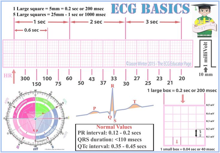
Ecg Nomal Waves And Segments Medstudent Ekg Basics Waves Segments Definitions Ecgeducator Segmentation Pr Interval Normal Ecg


Comments
Post a Comment